Diagnostic Testing
A Carotid ultrasound shows the amount of blood flow carotid arteries, the major blood vessels to the brain located on either side of the neck. With this imaging technique, your doctor can see if there is any narrowing of your carotid arteries because of cholesterol deposits or some other problem. Patients who had a stroke may have this test to evaluate the blood flow though the carotid arteries to determine cause of the first stroke and to determine the need for intervention to prevent further strokes.
What is a Carotid Ultrasound?
Carotid (ka-ROT-id) ultrasound is a painless and harmless test that uses high-frequency sound waves to create pictures of the insides of your carotid arteries.
You have two common carotid arteries, one on each side of your neck. They each divide into internal and external carotid arteries.
The internal carotid arteries supply oxygen-rich blood to your brain. The external carotid arteries supply oxygen-rich blood to your face, scalp, and neck.
Overview
Carotid ultrasound shows whether a waxy substance called plaque (plak) has built up in your carotid arteries. The buildup of plaque in the carotid arteries is called carotid artery disease.
Carotid Arteries
Figure A shows the location of the right carotid artery in the head and neck. Figure B is a cross-section of a normal carotid artery that has normal blood flow. Figure C shows a carotid artery that has plaque buildup and reduced blood flow.
Over time, plaque can harden or rupture (break open). Hardened plaque narrows the carotid arteries and reduces the flow of oxygen-rich blood to the brain.
If the plaque ruptures, a blood clot can form on its surface. A clot can mostly or completely block blood flow through a carotid artery, which can cause a stroke.
A piece of plaque or a blood clot also can break away from the wall of the carotid artery. The plaque or clot can travel through the bloodstream and get stuck in one of the brain’s smaller arteries. This can block blood flow in the artery and cause a stroke.
A standard carotid ultrasound shows the structure of your carotid arteries. Your carotid ultrasound test might include a Doppler ultrasound. Doppler ultrasound is a special test that shows the movement of blood through your blood vessels.
Your doctor might need results from both types of ultrasound to fully assess whether you have a blood flow problem in your carotid arteries.
Ultrasonography of the carotid arteries is frequently performed when there is a suspicion of carotid artery stenosis of occlusion. In this type of test an ultrasound probe covered with gel is held against the patients neck, and images are obtained of the carotid arteries. Measurements are made of the velocity of flow in these arteries which allows an estimation of the degree of vessel narrowing to be located
Carotid Stenosis
https://mayfieldclinic.com/pe-carotidstenosis.htm
Carotid artery screening is not recommended for asymptomatic stenosis
https://jamanetwork.com/journals/jama/fullarticle/2775715

Cerebral angiography has been used since the 1930s and still remains the “gold standard” for visualizing blood vessels of the brain. A small catheter is inserted in the groin, though which another thin catheter is passed over a flexible guidewire into each of the four arteries that carry blood to the brain. Then a dye containing iodine is injected and rapidly a sequence of x-rays is obtained as a blood carries the dye though the brain vessels. A computer removes the shadow of the bone and tissue so that only the blood vessels can be seen in fine detail.
Using catheters and this technique, stroke and many problems of brain blood vessels can now be treated such as brain aneurysms, arteriovenous malformations or dural arteriovenous fistulas.

CT Scan

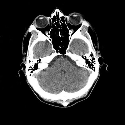
A computed tomography (CT) is the first diagnostic test used to diagnose a stroke. A CT scan uses X-rays to make detailed pictures of structures inside of the body. During the test, the patient lies on a table that is connected to the CT scanner. The scanner sends x-ray pulses though the body that takes pictures of thin slices of the brain, the images are saved to a computer where they can be viewed.
An iodine dye (contrast material) is often used to make the brain easier to see on the CT pictures. The dye may be used to check the blood flow, find tumors, and look for other problems. CT pictures may be taken before and after the dye is used.
A CT scan is an x-ray that is used to “see” inside the body. CT utilizes special x-ray equipment combined with computer technology. The patient lies down on a table, and is moved into the CT scanner for the testing. For CT scans of the head , most of the patient’s body stays outside the machine. The CT scanner takes “slices” of the tissue being examined. A CT scan of the brain takes less than 30 seconds to perform on a modern scanner. A CT scan is performed immediately on any new stroke patient. CT provides a picture of the brain which allows the differentiation of hemorrhagic from ischemic stroke. CT is valuable because it is fast, generally available at all times(there is a CT scanner in the ED), and can be performed on virtually any patient, including those with pacemakers and on ventilators. The disadvantage of CT scanning in acute stroke imaging is that it is relatively insensitive to early ischemic stroke changes even large strokes may not be visible immediately on CT, and it may miss strokes if they are small, or in such areas such as the brainstem which are difficult to image. A standard CT scan exposes the patient to a small amount of radiation.
CT angiography is a technique that can be used to demonstrate the appearance of blood vessels. This test is performed by giving the patient an infusion of intravenous contrast while simultaneously performing a CT scan which includes the area of interest. Contrast fills the blood vessels, and any vessel occlusion may be demonstrated. Special computer software may be used to represent these images in a more dramatic fashion. CT angiography is very often performed in a emergency room, where it is necessary to rapidly and accurately determine whether a major vascular occlusion has occurred, and help make decisions regarding therapy. CT angiography is an excellent test fir evaluation the carotid arteries in the neck. for demonstrate small vascular occlusions, and may be more accurate than MRA. It should be recognized that CT angiography carries the risk of intravenous contrast, and subjects the patient to significant radiation exposure
Echocardiogram
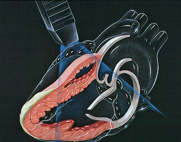
An echocardiogram is an ultrasound of the heart. In an echocardiogram, ultrasound gel is applied to the skin above the heart and a microscope-like instrument is applied to the chest wall. The technologist will move the instrument around in order to get the best possible view of the function of your heart. An echocardiogram enables a doctor to examine the working heart values, determines the size of your heart, and assess how well it is functioning. After a stroke this is important to determine if the heart was the source of the blood clot that caused the stroke.
The atria or fore chambers of the heart cannot be seen well on echocardiography though the chest wall. A more sensitive technique to see the back of the heart is called transesophageal echocardiogram (TEE). This is more often used in young patients with rarer causes of stroke such as a patent foramen ovale or atrial septal aneurysm.

MRI/MRA

Magnetic resonance imaging (MRI) is a technique that uses a magnetic field and radio waves to create detailed images of the organs and tissues within your body.
Most MRI machines are large, tube-shaped magnets. When you lie inside an MRI machine, the magnetic field temporarily aligns all the water molecules in your body. Radio waves cause these aligned particles to produce very faint signals, which are used to create cross-sectional MRI images – like slices in a loaf of bread. The MRI machine can combine these slices to produce 3-D images that may be viewed from many different angles.
A special sequence called “diffusion-weighted imaging” creates a special signal in brain tissue that has been damaged recently. This sequence is very important for the diagnosis of stroke.
MRI cannot be used in patients who have older aneurysm or surgical clips or in those who have pacemaker.
The same machine can also be used to obtain information about blood vessels in the brain. It can use the flow of blood to create this information or can be performed with a contrast called gadolinium.
MRI is another imaging technology used in stroke diagnosis. Exactly how MRI works is somewhat difficult to explain, but it uses magnetic fields and radio technology to produce images of the brain. MRI is capable of demonstrating both ischemic and hemorrhagic stroke, and may show ischemic stroke as early as minutes after it starts. It can demonstrate even very small strokes anywhere in the brain, including areas where CT may be negative. MRI uses no radiation. The disadvantages of MRI include the need for a patient to remain still for a period of 20 minutes in a relatively confined space, and the fact that it cannot be used in patients with pacemakers. MRI of the brain, when used for the diagnosis of stroke, is performed without IV contrast.
MRA, or magnetic resonance angiography, is a technique which also is able to demonstrate blood vessels. This technique uses a standard MRI machine with special software designed for this purpose. MRA of the head, which may demonstrate the intracranial circulation, is performed without contrast. MRA of the neck, which demonstrates the blood vessels leading from the heart to the brain is generally performed with contrast. The advantages of this test is that it does not expose the patient to radiation, and an image of the blood vessels inside the head may be obtained without contrast. The disadvantages of MRA is that it requires the patient to remain immobile for up to twenty minutes in a confined space. MRA may at times overestimate narrowing or occulsion of blood vessels.